Ventricular Tachycardia in Structural Hearts Disease

BACKGROUND

Ventricular tachycardia (VT) is an abnormal rapid heart rhythm originating from the lower pumping chambers of the heart (ventricles). The normal heart usually beats between 60 and 100 times per minute, with the atria contracting first, followed by the ventricles in a synchronized fashion. In VT, the ventricles beat at a rapid rate, typically from 120 to 300 beats per minute, and are no longer coordinated with the atria.
The controlled contraction of the ventricles is important for the heart to pump blood to the brain and the rest of the body and to maintain a normal blood pressure. Abnormal and fast rhythms from the ventricle may impair the ability of the pump to supply blood to the brain and the rest of the body as a result of the rapid rate and weak contractions.
  • Ventricular tachycardia (VT), an abnormal rapid heart rhythm originating from the lower pumping chambers of the heart with the ECG reading.
  • A dissection view of the heart showing the valves in the lower chambers.
  • A dissection view of the heart showing a focus on the RV outflow tract with the isolated RV outflow tract ectopics.
  • Ventricular tachycardia (VT), an abnormal rapid heart rhythm originating from the lower pumping chambers of the heart with the ECG reading.
  • A dissection view of the heart showing a focus on the RV outflow tract with the isolated RV outflow tract ectopics.
  • A dissection view of the heart showing a focus on the RV outflow tract with an ablation catheter.

SYMPTOMS

VT occurs most commonly in patients with weakened heart muscle (cardiomyopathy) or when scar tissue develops in the heart. In patients with coronary artery disease (blockage of blood vessels on the surface of the heart), this scar is the result of a prior heart attack (myocardial infarction) when the muscle dies as a result of a blockage in blood flow. Scar, or fibrosis, can interfere with the normal electrical impulse in the heart, leading to a short-circuiting of the rhythm, called reentry. VT can also occur in patients with normal hearts by a different mechanism whereby the electric conduction is overly excitable, like a muscle twitch. This may result in palpitations (a feeling of rapid or abnormal heart beat), dizziness, lightheadedness, or syncope (loss of consciousness). If the heart rate increases to more than 300 beats per minute and becomes totally uncoordinated, this is usually called ventricular fibrillation (VF), which will cause sudden cardiac death.
Sudden cardiac death causes about 450 000 fatalities each year in the United States alone. It is most commonly caused by VT deteriorating into VF, which is fatal within a few minutes if not defibrillated (shocked) back to a normal rhythm. Defibrillation may be accomplished by an automated external defibrillator or an implantable cardioverter-defibrillator (ICD). It is important to distinguish VT and VF, which are electric problems of the heart, from a heart attack, which is due to the sudden blockage of an artery. Heart attacks are treated with clot-busting drugs, balloon angioplasty, or stents. Sometimes, VT and VF are seen in that setting and are treated with electric shocks and drugs. The treatment of abnormal rhythms is discussed below.

TREATMENT

Treatment Options

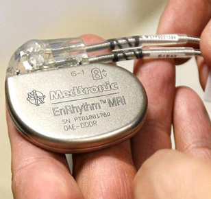
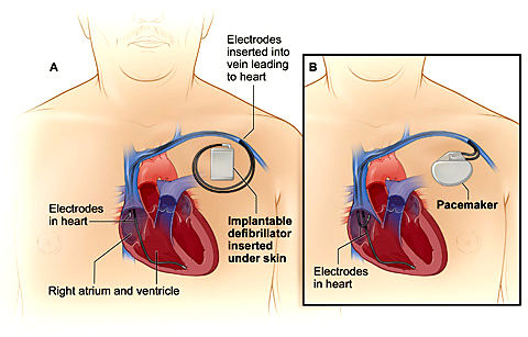
There are 3 treatment options for VT in patients with structural heart disease, although many patients require a combination: an ICD, antiarrhythmic medications, or catheter ablation. Many patients at risk for VT with structurally normal hearts are at risk of sudden cardiac death and as such are treated with an ICD. This is the most effective method of restoring a potentially life-threatening rhythm such as VT or VF back to a normal rhythm. However, an ICD does nothing to prevent the heart from going into VT. The ICD is a “safety net” and is like having an ambulance crew accompany you 24 hours a day. Please read our section on ICD.

Antiarrhythmic medications that modify the conduction of the electric impulse of the heart can be effective in suppressing VT. These medications can reduce the risk of recurrence by 75% but have potential side effects that include proarrhythmia, or worsening of the heart rhythm. For this reason, initiation of antiarrhythmic agents often requires close monitoring. Amiodarone, the most effective drug, has many side effects, which can involve toxicity to the vital organs like the liver, thyroid, lungs, eyes, and skin. Because of the discomfort associated with frequent ICD shocks and the side effects of antiarrhythmic drugs, catheter ablation is an important additional treatment option for many patients already using these therapies. The third treatment option is catheter ablation.

Catheter Ablation Therapy

Radiofrequency catheter ablation was first described 20 years ago, it has played an increasing role in the treatment of ventricular arrhythmias. Initially used in the treatment of patients with multiple ICD shocks for VT (VT storm), it is now used more frequently and earlier in the management of VT, particularly in centers with a high volume of patients and experience. Catheter ablation is an excellent choice for patients when medications are not effective, tolerated, or preferred.

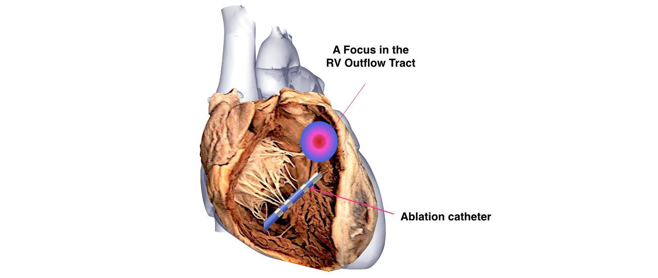
The aim of this procedure is to target the origin of the VT by placing a long, thin wire or catheter into the heart chambers through the veins of the leg. When areas that are critical to the VT circuit are identified, radiofrequency energy is applied to a small area (4 to 5 mm in diameter) to destroy the abnormal tissue. The number of burns required to treat the VT varies among patients. In patients with scar tissue in the heart, ablations may be performed within the scar and around its perimeter to cauterize or ablate the abnormal electric circuit responsible for the VT

-Senior Consultant Cardiologist & Electrophysiologist

-Chief, Cardiac Pacing and Arrhythmia Services

-Department of Cardiac Pacing and Electrophysiology

-Apollo Hospitals, Greams Road, Chennai.

© Arrhythmia Awareness Academy 2024 All Rights Reserved.
Designed by Digital SEO
chevron-down