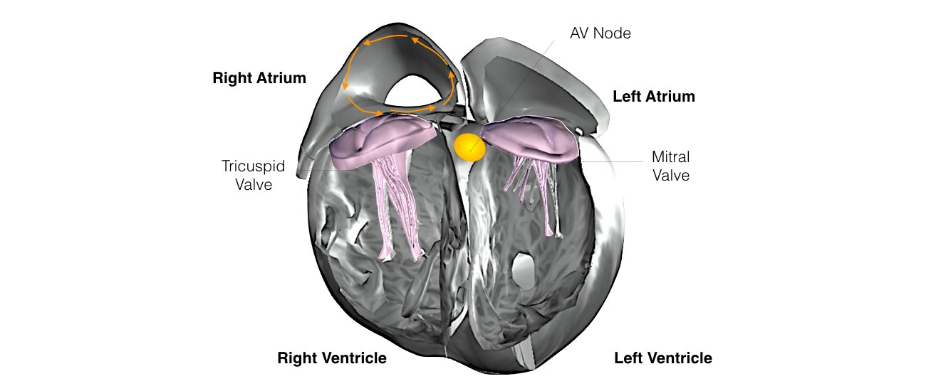
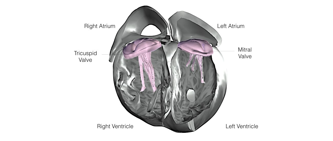
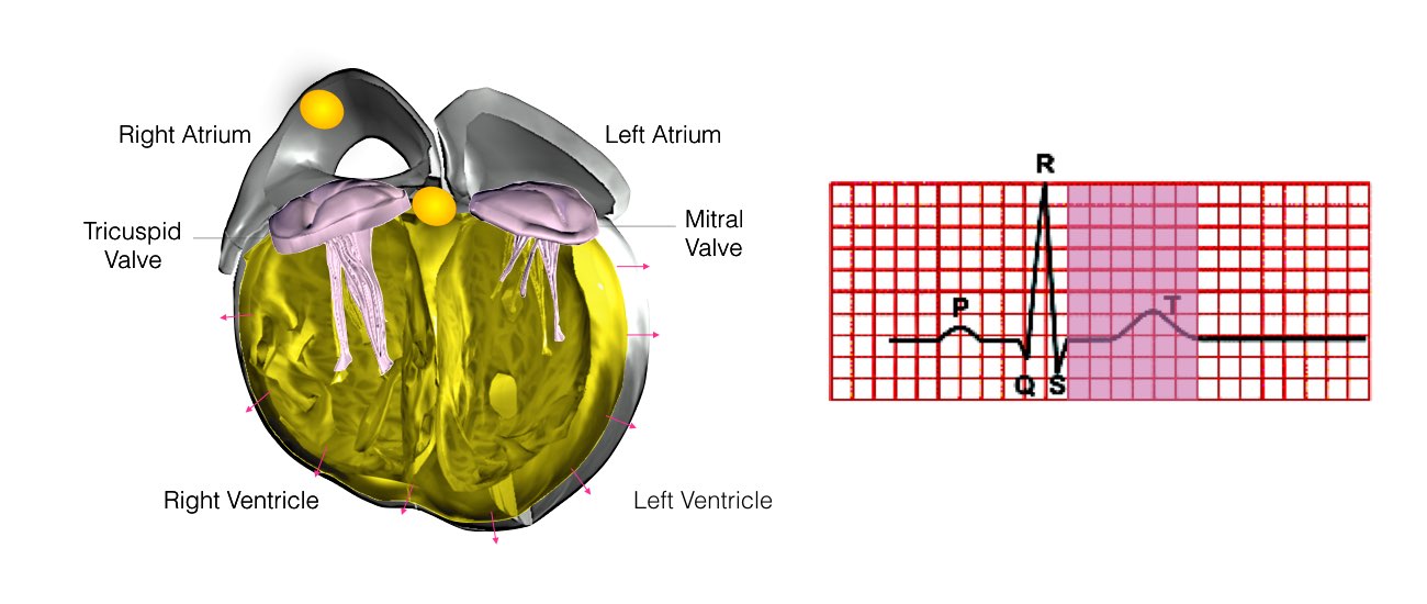
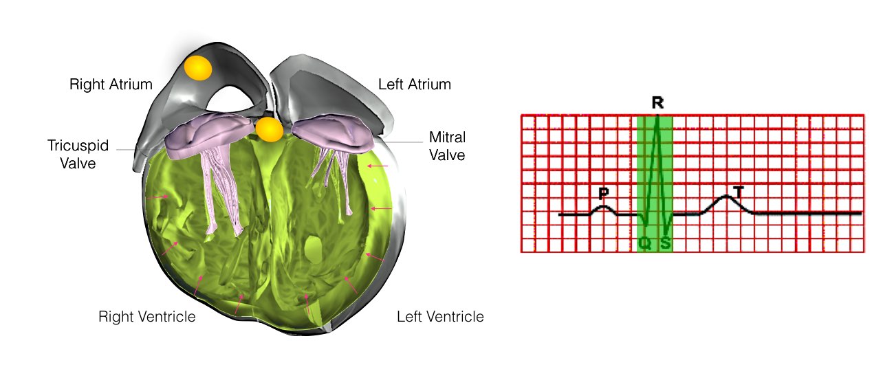
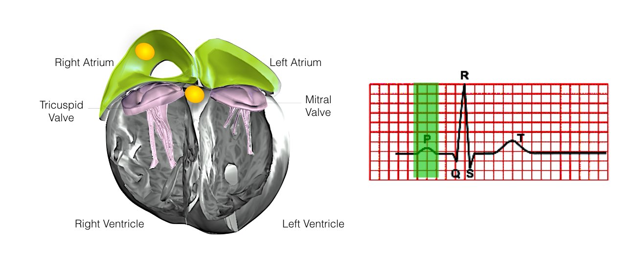
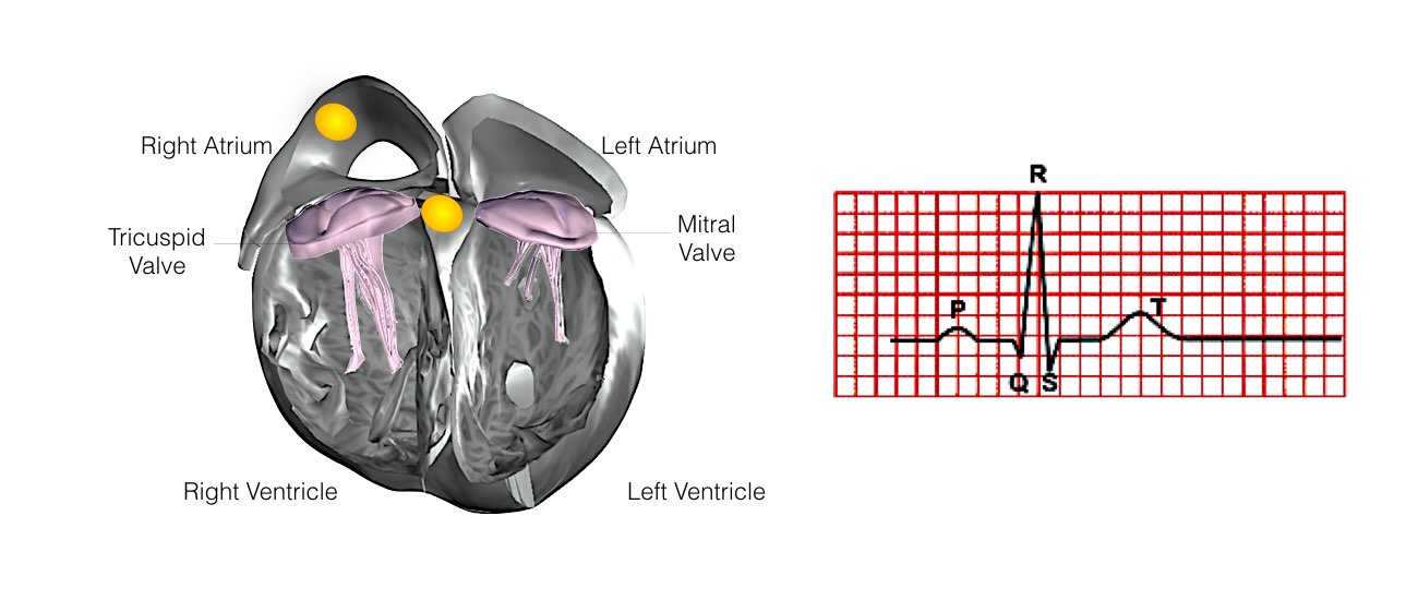
The heart is a pump responsible for maintaining blood supply to the body. It has four chambers. The two upper chambers (the right atrium and left atrium) are the chambers that receive blood as it returns from the body via the veins. The lower chambers (the right and left ventricle) are the chambers responsible for pumping the blood out to the body via the arteries. Like any pump, the heart has an electrical system that controls how it functions.
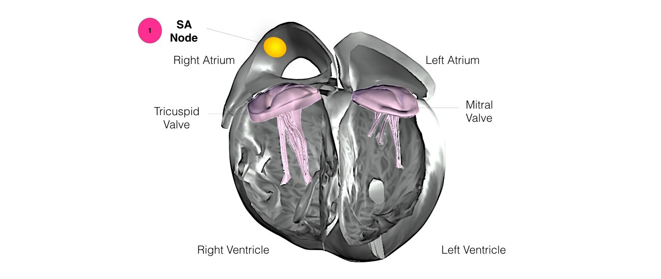
In order for the heart to do its work (pumping blood throughout the body), it needs a sort of spark plug or electrical impulse to generate a heartbeat. Normally, this electrical impulse begins in the upper right chamber of the heart (in the right atrium) in a place called the sino-atrial (SA) node. The SA node is the natural pacemaker of the heart. The SA node gives off electrical impulses to generate a heartbeat in the range of 60 to 100 times per minute. If you are exercising, doing strenuous work or are under stress, your heart rate will be faster. When you rest or sleep, your heart rate will slow down. If you take certain medications, your heart rate may be slower.
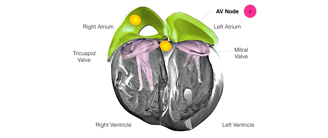
From the Sinus Node, the electrical impulse is relayed along the heart’s conduction system. It spreads throughout both the right and left atria, causing them to contract evenly. When the impulse spreads over the right atrium, it reaches the atrio-ventricular (AV) node. This is a very important structure in the heart because it is the only electrical connection between the top chambers and the bottom chambers. It is therefore the only way in which an electrical impulse can reach the pumping chambers (the ventricles). The impulse spreads through the AV node and down into the lower chambers or ventricles of the heart. This causes them to contract and pump blood to the lungs and body.
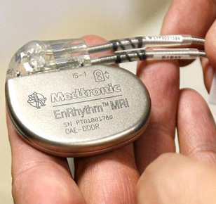
In some people, the electrical system of the heart may stop working properly. This can occur in a number of different ways. Sometimes the SA node fails to make enough impulses and the heart slows down and even pauses. This is sometimes called “sick sinus syndrome”. On other occasions, even though the SA node is making enough impulses, there are problems with either the AV Node, or the left and right bundle branches. When this happens, the impulses are not conducted down into the pumping chambers. This is termed “heart block” (This doesn’t mean there are blockages in arteries but rather in the electrical system). This will also cause the heart to slow down or pause. If your heart rate is too slow, you may need a pacemaker. These devices stimulate the heart to beat at a normal rate and pump more effectively.
When the heart slows down or pauses, symptoms may include tiredness, breathlessness or lightheadedness.
However, commonly when the heart pauses, you will experience dizziness or actually pass out and collapse. You may experience very little or no warning prior to collapsing
1. Single Chamber Pacemaker
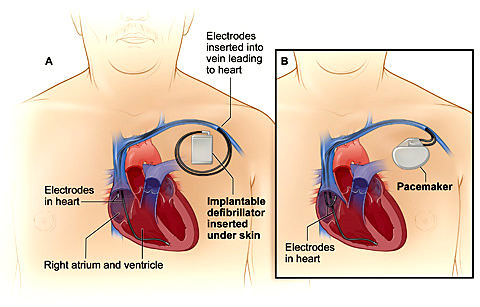
This type of pacemaker has one lead that connects the pulse generator to one chamber of your heart.
For most people, we use the single-chamber pacemaker to control heartbeat pacing by connecting the lead to your right ventricle (lower heart chamber). Depending on your symptoms and the type of pacing you need, we connect the lead to your right atrium (upper heart chamber) to stimulate the pacing in that chamber.
2. Dual Chamber Pacemaker
With two leads, this device connects to both chambers on the right side of your heart, the right atrium and the right ventricle. This pacemaker helps the two chambers work together, contracting and relaxing in the proper rhythm. The contractions allow blood to flow properly from the right atrium into the right ventricle.
Depending on the pacing needs of your heart, a dual-chamber device may be an appropriate option for you.
3. Physiological Pacing
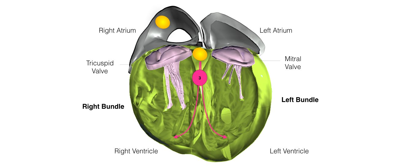
Physiological pacing is a new pacing technique where cardiac conduction system [HIS Bundle or Left Bundle] will be engaged while pacing the lower chamber of the heart. As pacing engages the normal electrical conduction system of the heart, thereby it prevents worsening of cardiac function and it is considered more physiological way of pacing the heart.
Traditional Pacemaker restores normal heartbeats in millions of people, but the widely using technique is placing the ventricular lead at lower Right Ventricle which increases risk of atrial fibrillation, heart failure and mortality.
Physiological pacing offers an elegant solution to avoid pacing-induced deterioration in cardiac function. Since activation occurs via the normal conduction system, it does not result in ventricular dyssynchrony and cardiac dysfunction.
