Electrophysiology Study
An EP Study is a specialized procedure conducted by a trained cardiac specialist, the Cardiac Electrophysiologist.
In this procedure, one or more thin, flexible wires, called catheters are inserted into a blood vessel (usually the groin) and guided into the heart. Each catheter has two or more electrodes to measure the heart’s electrical signals as they travel from one chamber to another. EP study is usually done under local anaesthesia.
An EP Study offers more detailed information about the heart’s electrical activity than many other noninvasive tests because electrodes are placed directly on heart tissue.
EP studies are done to diagnose your cardiac rhythm abnormality especially in a patient with palpitation or unexplained loss of consciousness, to help determine the best treatment.
EP study allows the Electrophysiologist to determine the specific location of an arrhythmia and, often, to correct it during the same procedure. This corrective treatment is considered a permanent cure; in many cases, the patient may not need to take heart medications.
Radiofrequency Catheter Ablation procedures for PSVTs
Radiofrequency Catheter Ablation – for patients with supraventricular tachycardias, atrial fibrillation and ventricular tachycardia
What Is Catheter Ablation?
Catheter ablation is a medical procedure used to treat some types of arrhythmia. An arrhythmia is a problem with the rate or rhythm of the heartbeat.
During catheter ablation, a long, thin, flexible tube is put into a blood vessel in your arm, groin (upper thigh), or neck. This tube is called an ablation catheter. It’s guided into your heart through the blood vessel.
A special machine, called a generator, sends energy through the ablation catheter to your heart. The energy modifies, via heating, or less commonly, freezing, small areas of heart tissue where abnormal heartbeats may cause an arrhythmia to start.
Radiofrequency (RF) energy usually is used for catheter ablation. This type of energy uses radiofrequency current to produce heat that modifies the heart tissue. Studies have shown that RF energy is safe and effective.
Who Needs Catheter Ablation?
To understand catheter ablation, it helps to understand how the heart works. The heart’s electrical system controls the rate and rhythm of your heartbeat.
Normally, with each heartbeat, an electrical signal spreads from the top of your heart to the bottom. As it travels, the electrical signal causes your heart to contract and pump blood. The process repeats with each new heartbeat.
A problem with any part of this process can cause an arrhythmia. Catheter ablation is one of several arrhythmia treatments. Your doctor may recommend it in several situations like :
- The medicines you take don’t control your arrhythmia.
- You can’t tolerate the medicines your doctor has prescribed for your arrhythmia due to side effects.
- You have abnormal electrical activity in your heart that raises your risk of ventricular tachycardia or fibrillation (a life-threatening arrhythmias) and sudden cardiac arrest.
Catheter ablation has some risks. Your doctor will explain the risks to you. In general, however, catheter ablation is a safe and effective curative procedure.
Before Catheter Ablation
Before you have catheter ablation, your doctor may review your medical history, do a physical exam, and recommend tests and procedures.
Your doctor will want to know about any medicines you’re taking. Some medicines can interfere with catheter ablation. If you take any of these medicines, your doctor may advise you to stop taking them before the procedure.
Your doctor also may ask whether you have diabetes, kidney disease, or other conditions. If so, your doctor may need to take extra steps during or after the procedure to help you avoid complications.
Before catheter ablation, you may have tests such as:
- EKG (electrocardiogram). This is a simple, painless test that records your heart’s electrical activity. The test shows how fast your heart is beating and its rhythm (steady or irregular). An EKG also records the strength and timing of electrical signals as they pass through each part of your heart.
- Echocardiography. This is a painless test that uses sound waves to create a moving picture of your heart. The test provides information about the size and shape of your heart and how well your heart chambers and valves are working.
- Stress testing. Some heart problems are easier to diagnose when your heart is working hard and beating fast. During stress testing, you exercise (or are given medicine if you’re unable to exercise) to make your heart work hard and beat fast while heart tests are done.
- Blood tests: Thyroid function tests may be done. An overactive or underactive thyroid gland may contribute to development of abnormal heart rhythms.
Less often, your doctor may recommend cardiac catheterization, coronary angiography or a test to rule out an overactive thyroid. (An arrhythmia can be a symptom of an untreated overactive thyroid.)
If you’re pregnant, let your doctor know before having catheter ablation. The procedure involves the use of radiation, which can harm a fetus. Talk with your doctor about whether you should have the procedure or wait. If you’re a woman of childbearing age, your doctor may recommend a pregnancy test before catheter ablation to make sure you’re not pregnant.
Once the procedure is scheduled, your doctor will tell you how to prepare for it. You’ll likely need to stop eating and drinking by midnight before the procedure. Your doctor will give you specific instructions.
Most people stay overnight following the ablation procedure. A few may require a longer than 1 day stay in the hospital.
During Catheter Ablation
Catheter ablation is done in a hospital. Doctors who do this procedure have special training in cardiac electrophysiology (the heart’s electrical system) and ablation (modification) of diseased heart tissue.
Before the Procedure
If you’re a woman of childbearing age, your doctor may recommend a pregnancy test before catheter ablation to make sure you’re not pregnant. The procedure involves the use of radiation, which can harm a fetus. If you’re pregnant, talk with your doctor about whether you should have the procedure or wait.
Before the procedure, you’ll be given medicine through an intravenous (IV) line inserted into a vein in your arm. The medicine will help you relax. It may make you sleepy. You’ll also be connected to several machines that will check your heart’s activity during the procedure.
Once you’re drowsy, your doctor will numb an area on your arm, groin (upper thigh), and/or neck. A needle will be used to make a small puncture through your skin and into a blood vessel. Your doctor will put a tapered tube called a sheath through this puncture site.
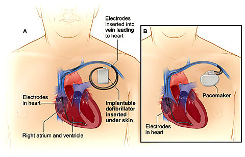
Your doctor will then put thin, flexible guide tubes and catheters (long, thin, flexible wires) into your blood vessel. These wires will be threaded through your blood vessels and into your heart. The wires have recording poles, called electrodes at the end. These are used to record electrical signals from inside the heart, similar to the way an ECG records signals from the body surface. The ablation catheter can record signals as well as be used to deliver energy through, to ablate, or modify the heart’s electrical tissue and cure the abnormal heart rhythm.
During the Procedure
Electrodes at the end of the catheter will be used to stimulate your heart and record its electrical activity. This will help your doctor learn where abnormal heartbeats are starting in your heart.
Your doctor will aim the tip of the catheter at the small area of heart tissue where the abnormal heartbeats are starting. A special machine will send energy through the catheter to create ablation lesions. In some cases, the abnormal heart rhythm may arise from a single point, or focus, while in other cases a circuit is involved and several ablation applications along one or more lines is needed. The ablation point or line(s) will modify the abnormal electrical tissue. This will stop abnormal electrical signals from traveling to the rest of the heart and causing arrhythmias.
What You May Feel
You will most likely be sleeping for most of the procedure. You may be awakened if it is difficult to start your abnormal rhythm in the sedated state, or for testing to see if the abnormal rhythm is gone after the ablation. You generally will not feel anything except possibly:
- A burning sensation when your doctor injects numbing medicine into the area where the catheter will be inserted
- Discomfort or burning in your chest when the energy is applied
- A faster heartbeat during studies that pinpoint the area(s) of your heart where abnormal heartbeats are starting
The procedure lasts 3 to 6 hours. When the procedure is done, your doctor will pull back the ablation catheter and take it out along with the sheath and guide wire.
The blood vessel puncture sites will be bandaged. Nurses will apply pressure to this site to help prevent major bleeding and to help the area begin to heal.
What To Expect After Catheter Ablation
After catheter ablation, you’ll be moved to a special care unit where you’ll lie still for 4 to 6 hours of recovery. Lying still prevents bleeding at the site where the ablation catheter was inserted.
While you’re in the special care unit, you’ll be connected to special devices that measure your heart’s electrical activity and blood pressure. Nurses will check these monitors regularly. Nurses also will check to make sure that you’re not bleeding from the catheter insertion site.
Going Home
You need to stay in the hospital for 1 or more days.
Before you go home, your doctor will tell you:
- Which medicines you need to take
- How much physical activity you can do
- How to care for the area where the catheter was inserted
- When to schedule followup care
Recovery and Recuperation
Recovery from catheter ablation usually is quick. You may feel stiff and achy from lying still for 4 to 6 hours after the procedure. You may receive pain medication for this following the procedure.
Also, a small bruise may form at the site where the catheter was inserted. The area may feel sore or tender for about a week. Most people are able to return to normal activities in a few days.
Your doctor will talk with you about signs and symptoms to watch for. Let your doctor know whether you have problems such as:
- A constant or large amount of bleeding at the catheter insertion site that you can’t stop with a small bandage
- Unusual pain, swelling, redness, or other signs of infection at or near the catheter insertion site
- Strong, rapid, or other irregular heartbeats
- Fainting
Follow-up care
The doctor will see you about 1 month after the procedure for follow-up. In the interim, if you note any unusual symptoms or have questions, please contact the office.
3D Mapping[CARTO 3] and Radiofrequency Catheter Ablation for Complex Atrial and Ventricular Arrhythmias like atrial fibrillation, atrial flutter, atrial and ventricular tachycardias.
3D mapping System is introduced for the benefit of the Cardiac Electrophysiologist in diagnosis and treatment of different complex cardiac arrhythmias.
The concept is to create the first 3-D image of the cardiac anatomy and electrical activity of the human heart with the best possible accuracy. This is done to localize the source of arrhythmia in the heart. The recent advances in this technology use a combination of magnetic and the electrical fields with special software to generate the visuals of the heart as 3 Dimensional images.
It uses special medical thin flexible insulated wires (better known as EP catheters), which allow the physicians to navigate, diagnose and deliver the desired therapy to the patient. The therapy is in the form of delivering Radiofrequency energy accurately through these catheters to the diseased tissue in the heart. This is one such therapy which is curative by nature.
This therapy is successful in the treatment of different cardiac arrhythmia disease states like Atrial Fibrillation or Ventricular Tachycardia or Atrial Flutter etc., if left untreated can lead to fatal incidences of stokes or sudden cardiac death loosing vital lives.
This procedure is done usually inside a cardiac catheterization lab [EP Lab] and the catheters are inserted into the patient body (through the groin or neck) and guided to the heart. This system also has the capability of interfacing with DICOM 3 format images obtained from CT and MRI facilities to assist the physician for better outcomes.